Pancreatic Islet Isolation by Miriam Ramírez-Domínguez
Author:Miriam Ramírez-Domínguez
Language: eng
Format: epub
Publisher: Springer International Publishing, Cham
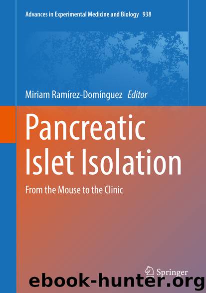
Fig. 5.3Collection of digested organ. (a) After stopping enzymatic activity, collected digested tissue is poured into a flask containing 1 L washing media; (b) Perfusing the chamber with washing media and collecting effluent from the chamber into 1 L bottles
Concentration and Purification of Islets
The dissociated tissue is collected in 250 ml conicals kept on ice, which are spun in refrigerated centrifuges for 1 min at 1000 rpm (Fig. 5.4a). After discarding the supernatants, pellets are pooled and the process is repeated until all the dissociated tissue is collected into one conical tube. The next step is islet purification . In general, we have less than 10 ml of digested tissue, and proceed to purify islets using a discontinuous gradient with Ficoll (polysucrose, Euro-Collins base, Mediatech), in a similar way as the one described for human islets [11, 20, 21]. Briefly, a sterile closed system is provided by using the COBE 2991 cell processor (COBE Laboratories, Inc., Lakewood, Colorado, USA), and ideally this procedure should to be done in a refrigerated cell processing room at 4 °C. The digested tissue is resuspended in 300 ml stock Ficoll (density 1.132 g/mL), loaded into a 600 ml transfer bag (Fig. 5.4b) and it is bottom loaded by gravity into the doughnut-shaped COBE bag (Fig. 5.4c). The discontinuous gradient is obtained by applying subsequently Ficoll solutions with density 1.108, 1.096 and 1.037 g/mL (75 mL each) and 50 ml Hanks at 2400 rpm. After a 3-min centrifugation, four fractions are collected into each of 250 ml conicals containing 100 ml of 10 % RPMI kept on ice: a 85 mL (layer #1), and 75 ml into each of the subsequent conicals (layers 2–4). The purest islets are generally found in layer #2, at the interface of 1.037/1.096 densities; less pure islets are found at the interface of 1.096/1.108 densities (layer 3); and the least pure islets are in layer 4, at the interface of 1.108/1.132 gradients. A sample from the tissue remaining in the COBE bag is taken to determine the presence of isolated islets or embedded islets after staining with dithizone. The conicals containing the islets are filled with 10 % RPMI and centrifuged at 1,500 rpm for 3 min at 4 °C. After removing the supernatant from each of the conicals, each pellet is resuspended in a final volume of 100 ml 10 % RPMI for layers 1 and 4 and 100 ml culture media at room temperature for layers 2 and 3. Figure 5.5 shows representative pictures of purified NHP islets stained with dithizone. They are shaped similar to human islets, with heterogeneous shapes and sizes. In general, islets obtained from the purest layer after purification are >90 % pure.
Fig. 5.4Concentration and purification of islets. (a) Digested tissue is collected into 250 ml conicals; (b) concentrated digest resuspended in stock Ficoll is poured into a 600 ml transfer bag; (c) contents of the transfer bag are emptied into the doughnut-shaped COBE bag located into the cell processor
Download
This site does not store any files on its server. We only index and link to content provided by other sites. Please contact the content providers to delete copyright contents if any and email us, we'll remove relevant links or contents immediately.
Whiskies Galore by Ian Buxton(42102)
Introduction to Aircraft Design (Cambridge Aerospace Series) by John P. Fielding(33192)
Small Unmanned Fixed-wing Aircraft Design by Andrew J. Keane Andras Sobester James P. Scanlan & András Sóbester & James P. Scanlan(32844)
Aircraft Design of WWII: A Sketchbook by Lockheed Aircraft Corporation(32341)
Craft Beer for the Homebrewer by Michael Agnew(18297)
Turbulence by E. J. Noyes(8143)
The Complete Stick Figure Physics Tutorials by Allen Sarah(7446)
The Institute by Stephen King(7109)
The Thirst by Nesbo Jo(7030)
Kaplan MCAT General Chemistry Review by Kaplan(7004)
Bad Blood by John Carreyrou(6685)
Modelling of Convective Heat and Mass Transfer in Rotating Flows by Igor V. Shevchuk(6504)
Weapons of Math Destruction by Cathy O'Neil(6390)
Learning SQL by Alan Beaulieu(6365)
Man-made Catastrophes and Risk Information Concealment by Dmitry Chernov & Didier Sornette(6158)
Permanent Record by Edward Snowden(5917)
Digital Minimalism by Cal Newport;(5853)
Life 3.0: Being Human in the Age of Artificial Intelligence by Tegmark Max(5636)
iGen by Jean M. Twenge(5475)
